Histology Reagents & Stains

DISPATCH TIME IN WEEKS: KIT DIMENSIONS: APPROXIMATE WEIGHT OF KIT: NO OF KITS IN SHIPPER: DIMENSION OF SHIPPER: APPROXIMATE WEIGHT OF SHIPPER: SAMPLES AVAILABILITY: PRODUCTION CAPACITY: |
PROCEDURE: 1. Spray Fixative on Slides. Drain excess. Dry in Air. RESULTS: |
CYTOCHROME STAIN WITH BUFFER
PROCEDURE: 1. Spray Fixative on Slides. Drain excess. Dry in Air. RESULTS: |
|
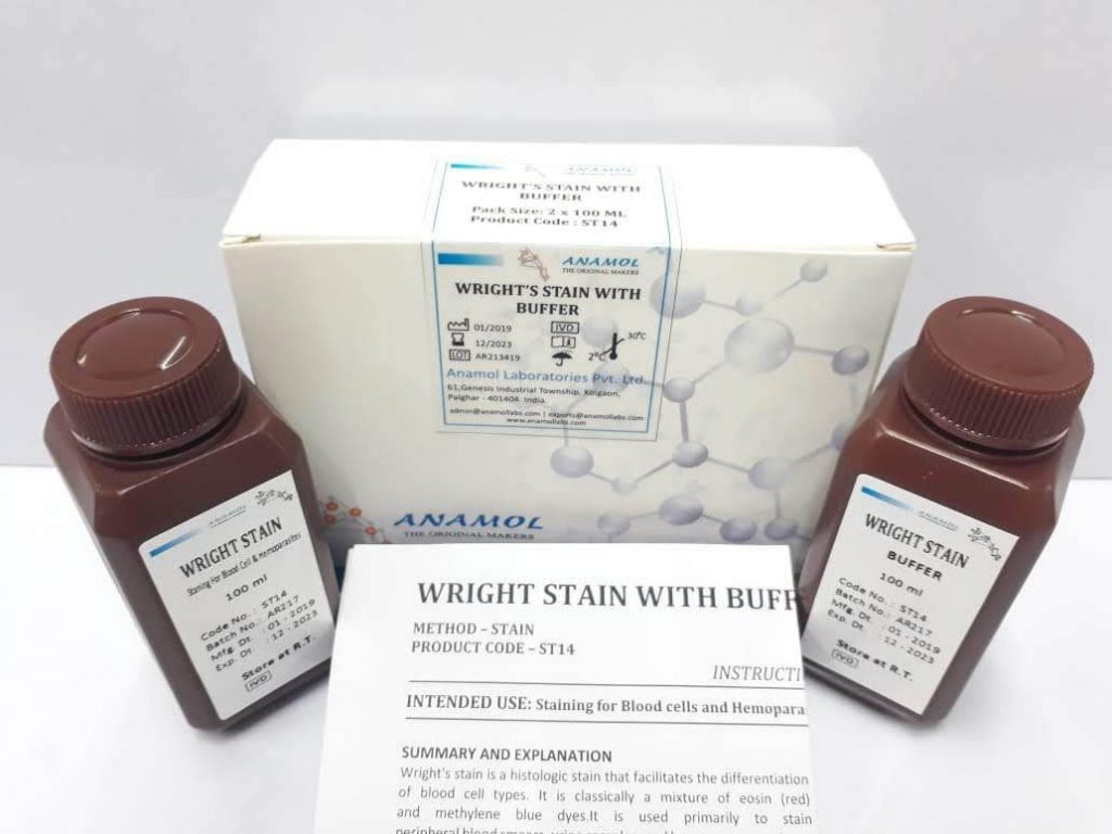
Wright’s Stain With Buffer
WRIGHT STAIN WITH BUFFER To detect intestinal protozoa Principle: Kit components: Physical Appearance: Pack Size: Storage Temperature: Shelf life: Signs of Deterioration
|
|
Availability:
In Stock.
Dispatch time in weeks:
2 to 4 weeks.
Kit dimensions:
2 x 50ml – 85x105x45 mm
4 x 50ml – 170x105x45 mm
4 x 250ml – 125x140X 125mm
Approximate weight of kit:
2 x 50 ml – 266 gms
4 x 50 ml – 493 gms
4 x 250ml – 1.1 kg
No of kits in shipper:
2 x 50 ml – 92 kits
4 x 50 ml – 46 kits
4 x250 ml – 12 kits
Dimension of shipper:
410x550x378 mm
Approximate weight of shipper:
2 x 50 ml – 26 kg
4 x 50 ml – 24 kg
4 x250 ml – 15 kg
Samples availability:
Yes
Production Capacity:
600 mn ml per annum.
PROCEDURE:
1. Spray Fixative on Slides. Drain excess. Dry in Air.
2. Pour Wright Stain on fixed slide. Keep it for 3 minutes.
3. Add Buffer. Keep it for 5 minutes.
4. Wash slide with distilled water.
5. Allow the smear to air dry.
6. Examine under oil immersion microscope.
RESULTS:
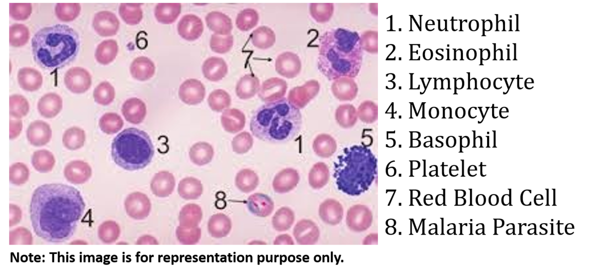
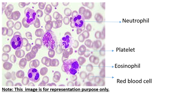
Neutrophils Dark Blue
Basophils Purple to Dark Blue
Lymphocytes Dark purple nuclei
Eosinophils Blue nuclei
Platelets Violet to Purple Granules
Dispatch time in weeks:
2 to 4 weeks.
Kit dimensions:
2 x 50ml – 85x105x45 mm
4 x 50ml – 170x105x45 mm
4 x 250ml – 125x140x125mm
Approximate weight of kit:
2 x 50 ml – 266 gms
4 x 50 ml – 493 gms
4 x 250ml – 1.1 kg
No of kits in shipper:
2 x 50 ml – 92 kits
4 x 50 ml – 46 kits
4 x250 ml – 12 kits
Dimension of shipper:
410x550x378 mm
Approximate weight of shipper:
2 x 50 ml – 26 kg
4 x 50 ml – 24 kg
4 x250 ml – 15 kg
Samples availability:
Yes
Production Capacity:
600 mn ml per annum.
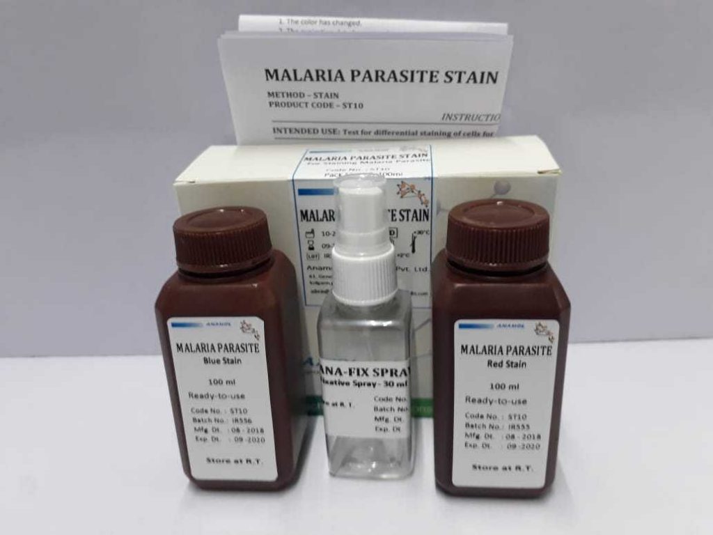
PROCEDURE:
1. Spray Fixative on Slides. Drain excess. Dry in Air.
2. Dip fixed slide in Red stain for 5-10 seconds.
3. Wash slide with distilled water.
4. Dip this slide in Blue stain for 45-50 seconds.
5. Wash slide with distilled water.
6. Allow the smear to air dry.
7. Examine under oil immersion microscope.
RESULTS:
Leucocyte Nuclei Dark Blue
Leucocyte Cytoplasm Light Blue
RBC Pale Reddish
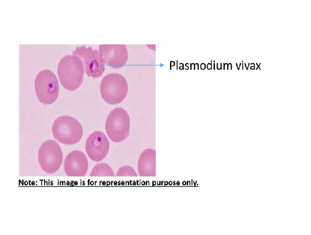
Malaria Parasite Varying shades of Blue
Platelets Pale or Dark Blue
Malaria Parasite Stain
MALARIA STAIN Intended use: Principle: Kit components: Physical Appearance: Pack Size: Shelf life: Signs of Deterioration: Availability:
|
|
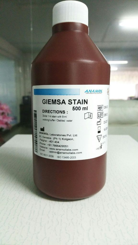
GIEMSA STAIN (READY TO USE)
INTENDED USE:
Test for differential staining of cells for detection of parasites by Giemsa stain.
PRINCIPLE:
Smears are fixed using the Giemsa Fixative. Slides are immersed in Giemsa Reagent to differentially stain specific cellular components. The cellular components stain either basophilic (blue) or eosinophilic (orange granules). The color intensity can be varied by adjusting the staining time.
KIT COMPONENTS:
Reagent bottles.
Kit insert.
PHYSICAL APPEARANCE:
Giemsa stain clear purple coloured liquid without any particles.
PACK SIZE:
1 x 100ml
1 x 500 ml
STORAGE TEMPERATURE:
Room Temperature.
SHELF LIFE:
60 months
SIGNS OF DETERIORATION:
1.Colour change
2.Growth
AVAILABILITY:
In Stock.
DISPATCH TIME IN WEEKS:
2 to 4 weeks.
KIT DIMENSIONS:
1 X100ml – 48x120x48mm.
1 x 500ml- 100x200X 100mm.
APPROX WEIGHT OF KIT:
1 x 100 ml – 130 gms.
1 x 500ml – 620 gms.
NO OF KITS IN SHIPPER:
1 x 100 ml – 200 kits.
1 x 500 ml – 18 kits.
DIMENSION OF SHIPPER:
410x550x378 mm.
APPROXIMATE WEIGHT OF SHIPPER:
1 x 100 ml – 27 kg.
1x 500 ml – 13 kg.
SAMPLES AVAILABILITY:
Yes.
PRODUCTION CAPACITY:
600 mn ml per annum.
PROCEDURE:
1. Pour Fixative on Slides. Drain excess. Dry in Air.
2. Pour Ready-To-Use Giemsa stain on the smear keep for 10 minutes.
3. Wash slide with distilled water.
4. Allow the smear to air dry.
5. Examine under oil immersion microscope
RESULTS
Nuclei Blue
Basophilic cytoplasmic Blue
Cellular Components
Neutrophilic granules Purple
Eosinophilic granules Orange
Lymphocytes Dark Blue
Mast cell granules Deep blue-violet
Nucleoli Blue-violet
Red cells Pink
Cytoplasm of mature monocytes Grey blue

Procedure: RESULTS: |
ALBERT′S STAIN A & B
Principle:
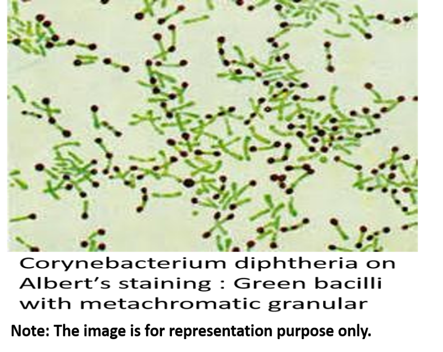
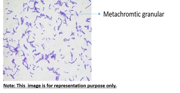
The granules stain purple-black against the light green counterstained cytoplasm. This helps to distinguish Corynebacterium diphtheria from most of the short nonpathogenic diphtheroides which lack granules.
Kit components:
Reagent bottles.
Kit insert.
Physical Appearance:
Alberts stain ‘A’ clear peacock blue coloured liquid without any particles.
Alberts stain ‘B’ clear amber coloured liquid without any particles.
Pack Size:
2 x 50 ml
4 x 50 ml
4 x 250 ml
Storage Temperature:
Room Temperature.
Shelf life:
60 months
Signs of Deterioration:
1. Colour change
2. Bacterial Growth
Availability:
In Stock.
Dispatch time in weeks:
2 to 4 weeks.
Kit dimensions:
2 x 50ml – 85x105x45 mm
4 x 50ml- 170x105x45 mm
4 x 250ml – 125x140x125mm
Approximate weight of kit:
2 x 50 ml – 266 gms
4 x 50 ml – 493 gms
4 x 250ml – 1.1 kg
No. of kits in shipper:
2 x 50 ml – 92 kits
4 x 50 ml – 46 kits
4 x250 ml – 12 kits
Dimension of shipper:
410x550x378 mm
Approximate weight of shipper:
2 x 50 ml – 26 kg
4 x 50 ml – 24 kg
4 x250 ml – 15 kg
Samples availability:
Yes
Production Capacity:
600 mn ml per annum.
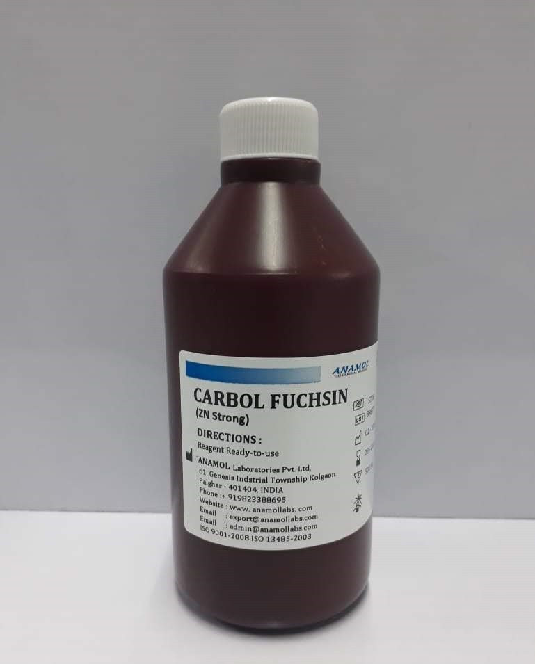
CARBOL FUSCHIN
Intended Use: Staining Solution for Microbes
Principle:
This differential staining technique is useful for identification of the tubercle bacillus, other Mycobacteria and Nocardia, which depends on the chemical composition of the bacterial cell wall. Because of the difficulty in staining these organisms with ordinary dyes, basic dyes in the presence of controlled amounts of acid are used. Generally, heat must be applied during the staining procedure, or wetting agents must be used, to aid dye penetration. Organisms exhibiting the property of acid-fastness, once stained, are not easily decolourized by alcohol.
Kit components:
Reagent bottles.
Kit insert.
Physical Appearance:
Carbol Fuschin stain – Clear wine-red coloured liquid without any particles.
Pack Size:
1 x 100ml.
1 x 500 ml.
Storage Temperature:
Room Temperature.
Shelf life:
60 months
Signs of Deterioration:
1. Colour change
2. Bacterial Growth
Availability:
In Stock.
Dispatch time in weeks:
2 to 4 weeks.
Kit dimensions:
1 X100ml – 48x120x48mm
1 x 500ml – 100x200X 100mm
Approximate weight of kit:
1 x 100 ml – 130 gms
1 x 500ml – 620 kg
No. of kits in shipper:
1 x 100 ml – 200 kits
1 x 500 ml – 18 kits
Dimension of shipper: 410x550x378 mm
Approximate weight of shipper:
1 x 100 ml – 27kg
1x 500 ml – 13 kg
Samples availability:
Yes
Production Capacity: |
PROCEDURE RESULTS |
PROCEDURE RESULTS |
|
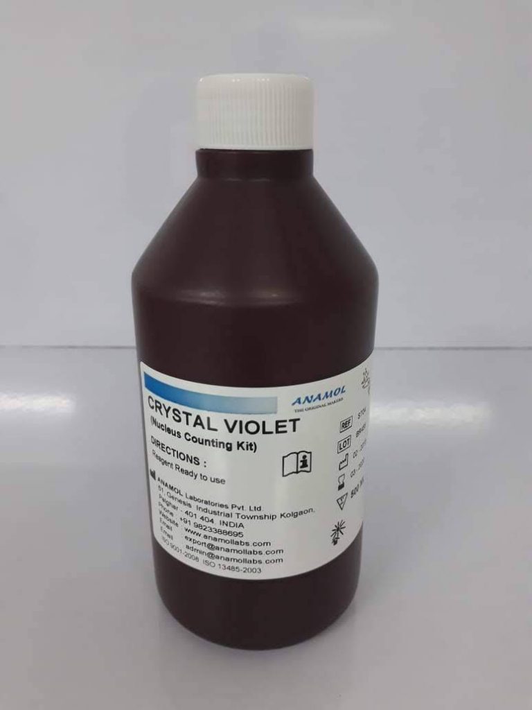
CRYSTAL VIOLET
Intended Use: Staining Solution for Bacteria.
Principle:
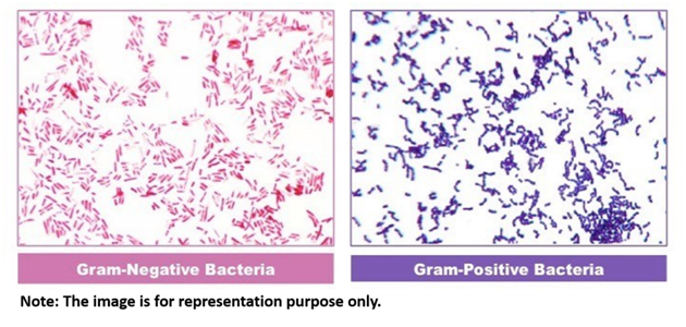
Crystal Violet is used as a simple stain where it can render the organisms violet. Besides this it can also be used in the Gram staining for distinguishing between gram-positive and gram-negative organisms.
Kit components:
Reagent bottles.
Kit insert.
Physical Appearance:
Crystal violet colored liquid without any particles.
Pack Size:
1 x 100ml
1 x 500 ml
Storage Temperature:
Room Temperature.
Shelf life:
60 months.
Signs of Deterioration:
1. Colour change.
2. Bacterial Growth.
Availability:
In Stock.
Dispatch time in weeks:
2 to 4 weeks.
Kit dimensions:
1 X100ml – 48x120x48mm
1 x 500ml – 100x200X 100mm
Approximate weight of kit:
1 x 100 ml – 130 gms
1 x 500ml – 620 kg
No. of kits in shipper:
1 x 100 ml – 200 kits
1 x 500 ml – 18 kits
Dimension of shipper: 410x550x378 mm
Approximate weight of shipper:
1 x 100 ml – 27kg
1x 500 ml – 13 kg
Samples availability:
Yes
Production Capacity:
600 mn ml per annum.
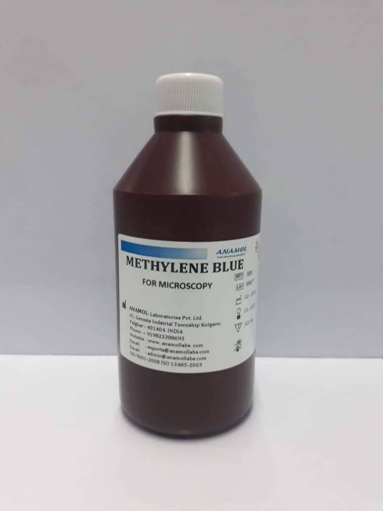
NEISSER′S METHYLENE BLUE
Staining Solution for Metachromatic staining.
Principle:
Well-developed granules of volutin (polyphosphate) may be seen in unstained wet preparations as round refractile bodies within the bacterial cytoplasm. With basic dye, they tend to stain more strongly than the rest of the bacterium, and with toluidine blue or methylene blue they stain metachromatically, a reddish purple colour. They are demonstrated most clearly by special methods, such as Albert’s and Neisser’s, which stain them dark purple but the remainder of the bacterium with a contrasting counterstain. The diphtheria bacillus gives its characteristic volutin-staining reactions best in a young culture (18-24 hours) on a blood or serum medium.
Kit components:
Reagent bottles
Kit insert.
Physical Appearance:
Neissers Methylene blue clear blue coloured liquid without any particles.
Pack Size:
1 x 100ml
1 x 500 ml
Storage Temperature:
Room Temperature.
Shelf life:
60 months
Signs of Deterioration
1. Colour change
2. Growth
Availability:
In Stock.
Dispatch time in weeks: Kit dimensions: Approximate weight of kit: No of kits in shipper: Dimension of shipper: Approximate weight of shipper: Samples availability: Production Capacity: PROCEDURE 9.Examine under oil immersion microscope. RESULTS |
|
PROCEDURE: RESULTS: |
|
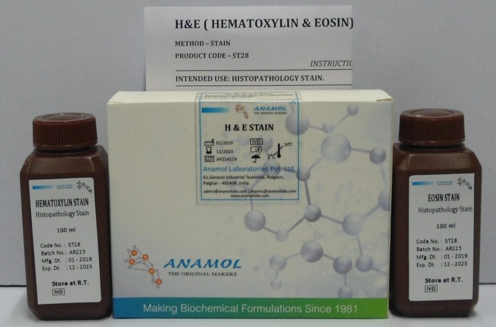
HEMATOXYLIN & EOSIN STAIN
HISTOPATHOLOGY STAIN.
Principle:
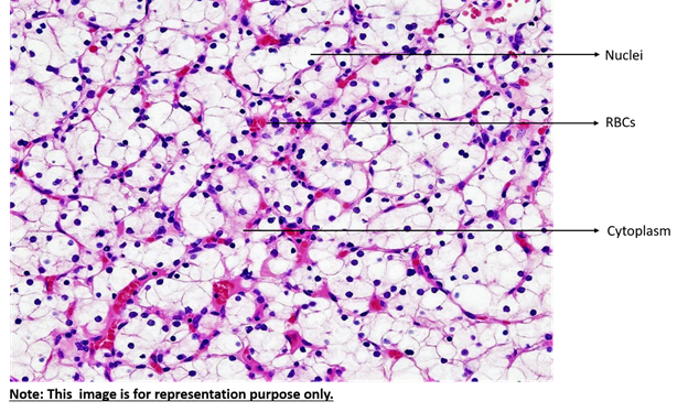
Alum acts as mordant and hematoxylin containing alum stains the nucleus light blue. This turns red in presence of acid, as differentiation is achieved by treating the tissue with acid solution. Bluing step converts the initial soluble red color within the nucleus to an insoluble blue color. The counterstaining is done by using eosin which imparts pink color to the cytoplasm.
Kit components:
Reagent bottles
Kit insert.
Physical Appearance:
Hematoxylin clear wine rad colored liquid without any particles.
Eosin clear reddish orange liquid without any particles.
Dehyrant clear colorless liquid without any particles.
Pack Size:
4 x 50 ml
4 x 250 ml
Storage Temperature:
Room Temperature.
Shelf life:
60 months
Signs of Deterioration
1. Colour change
2. Growth
Availability:
In Stock.
Dispatch time in weeks:
2 to 4 weeks.
Kit dimensions:
4 x 50ml- 170x105x45 mm
4 x 250ml – 125x140X 125mm
Approximate weight of kit:
4 x 50 ml – 493 gms
4 x 250ml – 1.1 kg
No of kits in shipper:
4 x 50 ml – 46 kits
4 x250 ml – 12 kits
Dimension of shipper:
410x550x378 mm
Approximate weight of shipper:
4 x 50 ml – 24 kg
4 x250 ml – 15 kg
Samples availability:
Yes
Production Capacity:
600 mn ml per annum.
